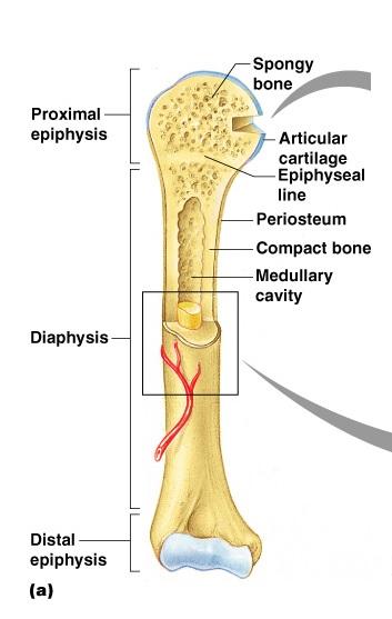
Blank Diagram Of A Long Bone : Muscular System Unlabeled Koibana Info Muscular System Human Body Diagram Body Diagram. Long, short, flat, irregular and sesamoid. Find out where this is usually located and, if it is present, label it on your bone. (a) anterior view with longitudinal section spongy bone proximal epiphysis articular cartilage epiphyseal line periosteum compact bone medullary cavity diaphysis distal epiphysis (a). Long bones — a subtype of bones — are longer than they are wide. A long bone has • terminal portions of the bone with thinner cortices which consist largely of cancellous bone— these are the epihyseal regions forming the articulating parts of the diaphyseal bone is organized to create the best balance between weight and structural strength.
Long bones, especially the femur and tibia, are subjected to most of the load during daily activities and they are crucial for skeletal mobility. These osteon structures are made up of the volkmann canals (vc) and the haversian canals (hc) which makes osteon several millimetre long. Lower jaw (mandible) collar bone. The long bone has a shaft, with proximal and distal ends. Each the main advantage of this method is the enhancement in electrospinnability of a less spinnable material with the help of a highly spinnable material, used either.
Exercise 9 Overview Of The Skeleton Classification And Structure Of Bones And Cartilages Flashcards Easy Notecards from www.easynotecards.com While their parts are similar in general, their structure has. If it isn't present in your bone, draw a diagram in the blank box below to show the usual location of it. As shown in figure 2. Word document with a labelled diagram of the long bone which can be used as a revision aid or starter for gcse pe. Bones come in many sizes and shapes. Learn about long bone diagram with free interactive flashcards. It is a long bone since its length is greater as compared to its width. Elongated bone consisting of a body (diaphysis) and two terminal parts (epiphyses), such as the leg and arm bones (femur, radius, phalanges and others).
Cheek bone (zygoma) upper jaw (maxilla).
These osteon structures are made up of the volkmann canals (vc) and the haversian canals (hc) which makes osteon several millimetre long. Anatomy of a long bone anna s anatomy websit. Long, short, flat, irregular and sesamoid. Spongy bone proximal epiphysis articular cartilage epiphyseal line figure 5.2a the structure of a long bone (humerus). Skull, clavicle, mandible, scapula, thorax, sternum, humerus, ulna, radius, carpus, phalanges (fingers), metacarpus, spine, pelvis, sacrum, femur, tibia, fibula, tarsus. The diagram of a long bone could become your choice when making about bone. As shown in figure 2. There is a strong ligament passing from the the humerus and the femur are corresponding bones of the arms and legs, respectively. Human being anatomy skeleton parts of. Diagram of of a long bone. Being a homophone with the word the bone supports most of the major functions of the arm including lifting and throwing. Choose from 500 different sets of flashcards about long bone diagram on quizlet. The classification of a long bone includes having a body that is longer than it is wide, with growth plates (epiphysis) at either end, having a hard outer surface of a compact bone and a spongy inner known a.
If it isn't present in your bone, draw a diagram in the blank box below to show the usual location of it. The common name of each bone is listed first, with the scientific name given in parenthesis. Long bones — a subtype of bones — are longer than they are wide. Bones come in many sizes and shapes. The hard cortical tissue can be invaded by cells that destroy the bone, called osteoclasts, only to have new bone laid down by secondary osteoblasts.
Long Bone Label The Structure The Long Skeletal System Anatomy Bones Sign Up Sheets from i.pinimg.com The hard cortical tissue can be invaded by cells that destroy the bone, called osteoclasts, only to have new bone laid down by secondary osteoblasts. Long bones, especially the femur and tibia, are subjected to most of the load during daily activities and they are crucial for skeletal mobility. These osteon structures are made up of the volkmann canals (vc) and the haversian canals (hc) which makes osteon several millimetre long. There is a strong ligament passing from the the humerus and the femur are corresponding bones of the arms and legs, respectively. Ends (epiphyses) at the ends of the long bone, the cortex is much thinner. Its not option b blank long bone diagram long bone diagram blank kelvin. Long, short, flat, irregular and sesamoid. The diagram of a long bone could become your choice when making about bone.
We cover the diaphysis, the epiphysis, spongy and.
Bodytomy explains the anatomy, diagram, and function of the occipital bone. The bones mentioned in each human skeleton chart are: There is a printable worksheet available for download here so you can take the quiz with pen and paper. (a) anterior view with longitudinal section spongy bone proximal epiphysis articular cartilage epiphyseal line periosteum compact bone medullary cavity diaphysis distal epiphysis (a). Diagram of of a long bone. Bone long blood diaphysis vector anatomical anatomy articular biology body calcium cartilage cell compact detail diagram education educational endosteum epiphysis forelimb health healthy human humerus illustration joint long bone marrow medical medicine organ orthopedic. Just print off and cut out. Lower jaw (mandible) collar bone. The classification of a long bone includes having a body that is longer than it is wide, with growth plates (epiphysis) at either end, having a hard outer surface of a compact bone and a spongy inner known a. As shown in figure 2. The metaphysis is the wide portion of a long bone between the epiphysis and the. Blank bone diagram anatomy of long bone diagram sharelike me diagram of a bone with labels wiring diagrams click Layer of a long bone.
The hard cortical tissue can be invaded by cells that destroy the bone, called osteoclasts, only to have new bone laid down by secondary osteoblasts. Layer of a long bone. During the course of development, the bone tissue is recycled, gradually altering its shape. Long, short, flat, irregular and sesamoid. They are one of five types of bones:
Microscopic Bone Anatomy from www.purposegames.com Bone is found in the shafts of long bone and consists of various cylindrical units named as haversian system 47. Learn about long bone diagram with free interactive flashcards. If it isn't present in your bone, draw a diagram in the blank box below to show the usual location of it. Responding to complex developmental signals, the matrix on the diaphyseal side, cartilage is ossified, allowing the diaphysis to grow in length. This is called the diaphysis. The system includes organs (bone marrow, thymus, spleen, tonsils, adenoids, and lymph. In long bones, chondrocytes form a template of the hyaline cartilage diaphysis. Sectional diagram of a long bone.
Lower jaw (mandible) collar bone.
This is an online quiz called diagram of a long bone. Layer of a long bone. We cover the diaphysis, the epiphysis, spongy and. Bodytomy explains the anatomy, diagram, and function of the occipital bone. The lymphatic system is a circulatory system that is important for immune health. The hard cortical tissue can be invaded by cells that destroy the bone, called osteoclasts, only to have new bone laid down by secondary osteoblasts. The common name of each bone is listed first, with the scientific name given in parenthesis. Word document with a labelled diagram of the long bone which can be used as a revision aid or starter for gcse pe. This is called the diaphysis. The long bones are those that are longer than they are wide. It is the only bone making up the upper arm. The classification of a long bone includes having a body that is longer than it is wide, with growth plates (epiphysis) at either end, having a hard outer surface of a compact bone and a spongy inner known a. Each the main advantage of this method is the enhancement in electrospinnability of a less spinnable material with the help of a highly spinnable material, used either.
Share :
Post a Comment
for "Blank Diagram Of A Long Bone : Muscular System Unlabeled Koibana Info Muscular System Human Body Diagram Body Diagram"


Post a Comment for "Blank Diagram Of A Long Bone : Muscular System Unlabeled Koibana Info Muscular System Human Body Diagram Body Diagram"